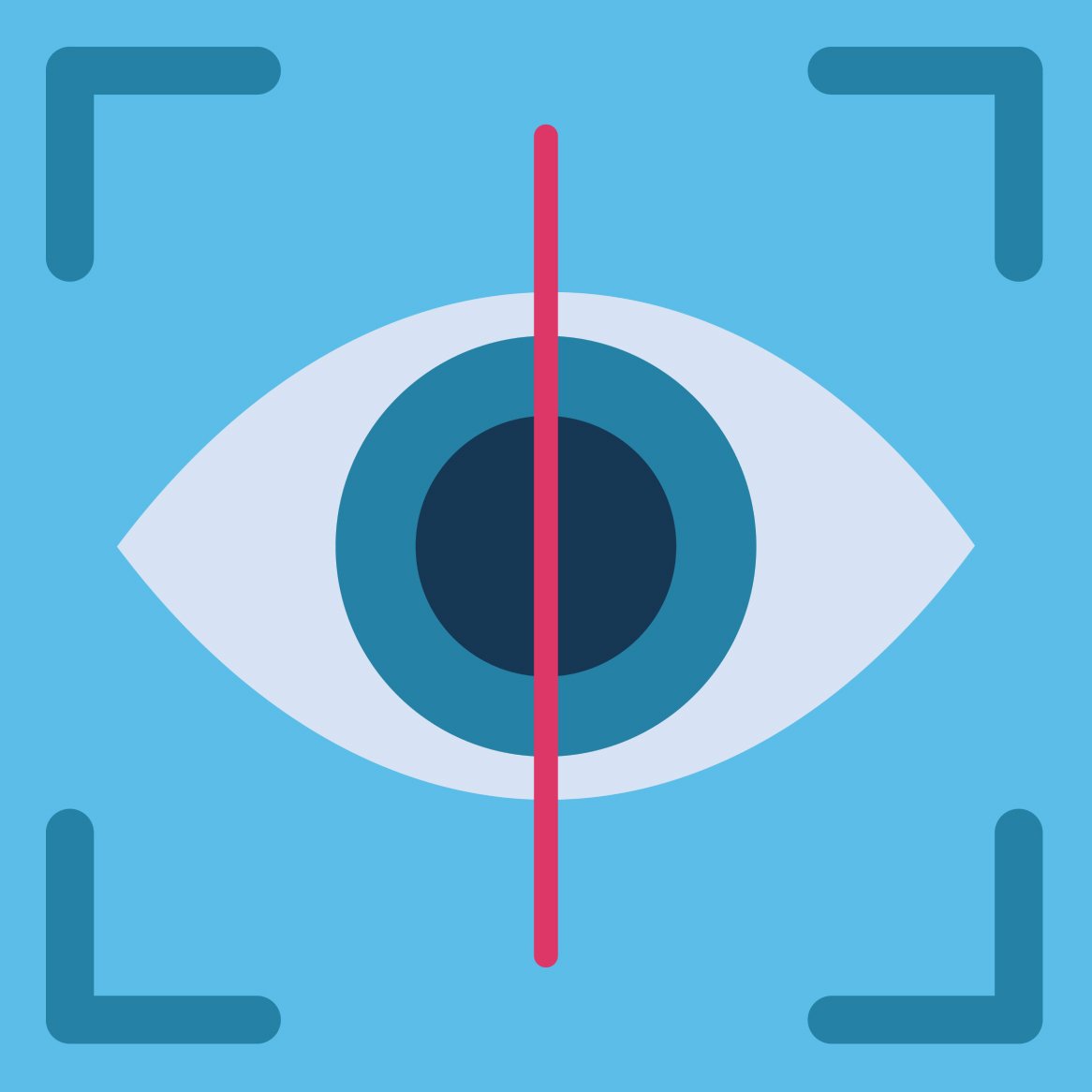
Retinal imaging uses digital photography to help eye doctors check your eye health—and aspects of your overall health—during a routine eye exam.
Retinal imaging systems take high-resolution digital pictures of the back of your eye, including the retina, the optic disc, the optic nerve, and blood vessels. These images can be used as a diagnostic tool to help detect vision- or life-threatening disorders and diseases early, often before symptoms appear. And getting early treatment can be essential in combating these issues or minimizing their effects.
In this article, we’ll go into more detail about how retinal imaging works. We’ll also go over the finer points of how it can benefit patients and help maintain their health.
What Is Retinal Imaging?
Retinal imaging can be an important part of your overall healthcare. It works by taking special scans of your eyes. The images left behind are retrieved and then used to create detailed pictures of the back of your eye.
One of the primary benefits of retinal imaging is that it’s noninvasive. This allows eyecare professionals to obtain detailed information as safely as possible.
What Does Retinal Imaging Test For?
Optometrists and ophthalmologists use retinal imaging to check if certain eye conditions (including some that could lead to vision loss) may be present or developing, like:
- Diabetic retinopathy
- Glaucoma
- Macular degeneration
- Retinal detachment
Retinal imaging can also aid in the early detection of other health problems like stroke, cardiovascular disease, hypertension (high blood pressure), and some types of cancer.
Besides providing a way to help assist in the diagnosis of certain health issues in real time, retinal imaging gives doctors a record of what your eye looks like at different points in your life. Comparing each year’s photos can assist doctors in detecting subtle eye changes that could signal an emerging problem.
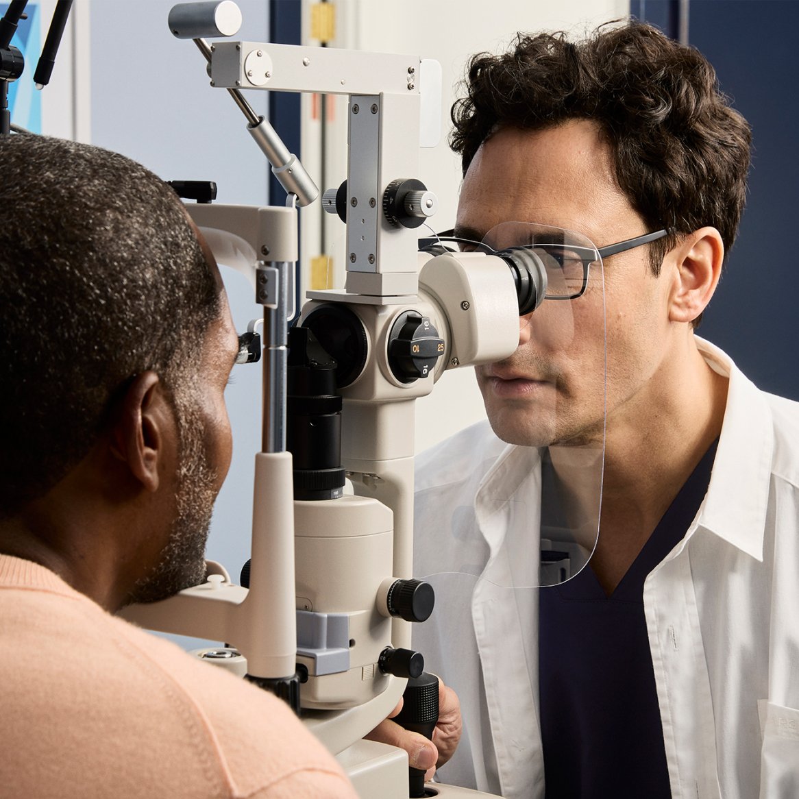
What Happens During a Retinal Exam?
Don’t worry—no one’s sticking a camera in your head, and nothing touches your eye at any time. Retinal imaging is a painless (dare we say comfortable?) and speedy technique.
A retinal exam is just one part of a comprehensive eye exam. And if you’re getting a retinal scan in place of pupil dilation (we’ll talk more about this in a bit), it can shorten the time your eye exam takes. Let’s look at how it happens:
- Gaze into the device: Your doctor will direct you to look into the machine one eye at a time. It might seem a bit like looking through a peephole.
- Flash: When you see a brief flash of light, you’ll know the image has been taken.
- Behold your retina: The digital images are immediately ready for you and your doctor to view.